High-throughput, low-cost FLASH: irradiation of Drosophila melanogaster with low-Energy X-rays using time structures spanning ConvDR and UHDR

This article explores the potential of using low-energy X-rays to deliver ultrahigh dose-rate (UHDR) FLASH radiotherapy using Drosophila melanogaster as a model. For this they have compared the effects of UHDR (210 Gy/s) and conventional dose rates (0.2–0.4 Gy/s) on the eclosion and lifespan of fly larvae. The results showed that larvae treated with UHDR had higher survival rates and longer lifespans, particularly at intermediate doses, indicating a normal tissue-sparing FLASH effect.

The Medscint scintillation dosimetry detector was used to measure the response to X-rays at a very high sampling rate to confirm the time structure of the delivered radiation (i.e. the pulse width and inter-pulse spacing). Along with film measurements, they also confirmed that the doses delivered with UHDR and CONV agreed within 0.1%.

Journal of Radiation Research
Alexander Hart (1), Jan P Dudzic (1), Jameson W Clarke (1), Jonathan Eby (2), Steve J Perlman (1), Magdalena Bazalova-Carter (1) | 1. University of Victoria, BC – CANADA, 2. University of Toronto, ON – CANADA

Field output correction factors using a fully characterized plastic scintillation detector (HYPERSCINT RP-200)

As small fields become increasingly important in radiation therapy, accurate dosimetry is essential for ensuring precise dose calculation and treatment optimization. Despite the availability of small volume detectors, small field dosimetry remains challenging. The new plastic scintillation detector (PSD) from the HYPERCINT RP-200 platform from Medscint offers a promising solution with minimal correction requirements for small field measurements.

This study focused on characterizing the field output correction factors of the PSD across a wide range of field sizes and demonstrating its potential for determining correction factors for other small field detectors. Monte Carlo simulations and experimental comparisons were used to assess the system’s performance. The PSD exhibited near-unity correction factors (1.002 to 0.999) across field sizes between 0.6×0.6 cm² and 30×30 cm², with an impressive total uncertainty of 0.5%.

The PSD is shown to be a highly accurate and reliable detector for small field dosimetry, and it can also be used to determine correction factors for other dosimeters with great precision.

PREPRINT
Luc Gingras (1,2), Yunuen Cervantes (1,2,3), Frederic Beaulieu (1,2), Magali Besnier (1,2), Benjamin Coté (4), Simon Lambert-Girard (4), Danahé LeBlanc (4), Yoan LeChasseur (4), François Therriault-Proulx (4), Luc Beaulieu (1,2,3), Louis Archambault (1,2,3) | 1. CHU de Québec–Université Laval, Québec – Canada, 2. Centre de recherche du CHU de Québec, Québec – Canada, 3. Université Laval, Québec – Canada, 4. Medscint, Québec – Canada

Development and first implementation of a novel multi-modality cardiac motion and dosimetry phantom for radiotherapy applications

Magnetic resonance guided radiation therapy (MRgRT) for real-time gating around the heart for treating ventricular tachycardia (VT) are rapidly advancing. A novel, multi-modality modular heart phantom was developed and utilized in gated radiotherapy experiments on a 0.35 T MR-linac. This phantom can simulate cardiac, cardio-respiratory, and respiratory motions, and perform dosimetric evaluations using ionization chamber and plastic scintillation detectors (PSD from MEDSCINT) configurations.

Due to their small sensitive volumes, time-resolved PSDs are effective for low-amplitude/high-frequency movements and multi-point data acquisition, enhancing dosimetric capabilities. This advancement in VT planning and delivery illustrates the phantom’s potential to meet the growing demands of cardiac applications in radiotherapy.

MEDICAL PHYSICS
Kenneth W. Gregg (1,2), Chase Ruff (1,2), Grant Koenig (3), Kalin I. Penev (3), Andrew Shepard (1), Grace Kreissler (4), Margo Amatuzio (4), Cameron Owens (4), Prashant Nagpal (5), Carri K. Glide-Hurst (1,2) | 1. Department of Human Oncology, University of Wisconsin–Madison, Madison, Wisconsin, USA, 2. Department of Medical Physics, University of Wisconsin–Madison, Madison, Wisconsin, USA, 3. Modus Medical Devices, Inc. (IBA QUASAR),London, Ontario, Canada, 4. Department of Biomedical Engineering, University of Wisconsin–Madison, Madison, Wisconsin, USA, 5. Department of Radiology, University of Wisconsin–Madison, Madison, Wisconsin, USA

Characterization of a multi-point scintillation dosimetry research platform for a low-field MR-Linac

Plastic scintillation detectors (PSDs) are attractive for enhancing MRI-guided radiation therapy (MRgRT). A study evaluating the HYPERSCINT RP-200, a multi-probe PSD system, demonstrated excellent repeatability and minimal deviation in performance metrics such as detector response and percent depth dose (PDD). PSDs maintained consistent linearity across a broad range of monitor units and showcased high accuracy in gating experiments (ex. gating experiments where 400 cGy were delivered to isocenter : < 0.8 cGy variation for central axis measures and < 0.7 cGy for the gradient sampled region). These results highlight PSDs' huge potential in improving the precision and reliability of MRgRT, especially in complex real-time applications.

MEDICAL PHYSICS
Jennie Crosby (1), Chase Ruff (1), Ken Gregg (1), Jonathan Turcotte (2), Carri Glide-Hurst (1) | 1. Department of Human Oncology, University of Wisconsin-Madison, Madison, Wisconsin, USA, 2. Medscint, Québec, Quebec, Canada

MLC tracking and dose accumulation validation on the MR-linac using a real-time deformable dosimeter

Online MRI on the MR-linac captures detailed anatomical movements, improving real-time radiotherapy adaptations. However, the lack of a suitable MRI-compatible phantom hinders workflow validation. This study introduces a deformable phantom with integrated real-time scintillation dosimeters, validating accuracy in MLC tracking and dose accumulation using the ELEKTA Unity MR-linac.

This study demonstrates the vast potential of this novel prototype deformable phantom with integrated PSDs for real-time dosimetry measurements on an MR-linac.

2024 ESTRO Annual Congress
Madelon van den Dobbelsteen (1), Pim T.S. Borman (1), Laurie J.M. de Vries (1), Sara L. Hackett (1), Kalin Penev (1), Rocco Flores (2), Stephanie Smith (2), Yoan LeChasseur (3), Simon Lambert-Girard (3), Benjamin Côté (3) , Peter L. Woodhead (1)(4), Lando S. Bosma (1), Cornel Zachiu (1), Bas W. Raaymakers (1), Martin F. Fast (1) | 1 University Medical Center Utrecht, Radiotherapy, Utrecht, Netherlands., 2 IBA QUASAR, Modus Medical Devices Inc. London ON, Canada. , 3 Medscint, -, Quebec City, Canada. , 4 Elekta AB, -, Stockholm, Sweden.

Experimental dosimetric verification of the intra-fraction drift correction on the 1.5 T MR-linac

MRI-guided online adaptive treatments can improve tumor targeting by adjusting treatment plans in real-time based on cine MR-scans. And to correct the intra-fraction motion, Elekta AB introduced the intra-fraction drift correction (IDC) functionality for the 1.5 T Unity MR-linac.

The IDC is a valuable functionality for fast intra-fraction adaptations and this research experimentally verifies the geometric and dosimetric accuracy of the IDC process using film, scintillation, and diode dosimetry.

ESTRO 2024 Annual Congress
Madelon van den Dobbelsteen, Sara L. Hackett, Stijn Oolbekkink, Bram van Asselen, Prescilla Uijtewaal, Martin F. Fast, Bas W. Raaymakers | University Medical Center Utrecht, Radiotherapy, Utrecht, Netherlands

Evaluation of the HYPERSCINT scintillation dosimetry platform for small-field characterization of a Leksell GAMMA KNIFE

The performance of the HYPERSCINT RP-200 with the 0.5mm x 0.5mm detector was evaluated for the characterization of small radiation fields administered using a Leksell Gamma Knife Perfexion radiosurgery device. Overall, our results show that the detector response was in close agreement with Gamma Knife Monte Carlo reference data and film measurements. Based on the obtained results, the plastic scintillation detector shows the potential for rapid validation of output factors and validation of film measurements as well its use in challenging small-field situations encountered with the Gamma Knife.

ISRS 2024 – New York
Mathieu GUILLOT (1), Patrick DELAGE (1), Vincent HUBERT-TREMBLAY (1), Francois THERRIAULT-PROULX (2), Danahé LEBLANC (2) | CHUS – Sherbrooke, Canada, Medscint – Québec, Canada

Development of End-to-End Preclinical Treatment Verification Procedures, Traceable to NPL Air Kerma Primary Standard

Dosimetry audits are an important tool to improve quality of reported results and to support standardization of preclinical radiation research. This work presents how the combination of passive and active detectors, such as the real-time HYPERSCINT scintillation dosimetry solution, with anatomically correct mouse phantoms are adequate for the development of End-to-End dosimetry audits for the independent verification of preclinical radiation treatments.

The traceability of the detectors’ calibration to primary standards strengthens the dosimetry chain in the validation of preclinical plans, and it is consistent with the current practice for dose traceability of clinical radiotherapy treatments. Their implementation at national and regional levels could lead to databases of anonymised records, which will positively impact the dissemination of best practices and sharing of validated results.

6th Conference on small animal precision image-guided radiotherapy
Ileana Silvestre Patallo (1), Rebecca Carter (2)(3), Andrew Nisbet (2), Anna Subiel (1), Giuseppe Schettino (1) | 1. National Physical Laboratory, UK, 2. University College London, UK, 3. Cancer Institut, UK

Characterization of a novel time-resolved, real-time scintillation dosimetry system (HYPERSCINT RP-FLASH) for ultra-high dose rate radiation therapy applications

This study evaluates a novel scintillation dosimetry solution developed by Medscint for ultra-high dose rate (UHDR) radiotherapy, the HYPERSCINT RP-FLASH. The system was tested on an UHDR electron beamline, demonstrating dose linearity and independence from dose rate (1.8–1341 Gy/s) and dose per pulse (0.005–7.68 Gy) within ±3% tolerance. The system accurately measured doses per pulse up to 120 Hz.

With daily calibrations and specific correction factors, the system provides real-time, millisecond-resolved dosimetric measurements for pulsed conventional and UHDR beams, showing promise for applications in FLASH-RT.

PREPRINT
Alexander Baikalov (1,2,3), Daline Tho (1), Kevin Liu (1,4), Stefan Bartzsch (2,3), Sam Beddar (1,4), Emil Schüler (1,4) | 1. University of Texas MD Anderson Cancer Center, Houston, TX – USA, 2. Technical University of Munich – Germany, 3. German Research Center for Environmental Health, Neuherberg – Germany, 4. The University of Texas MD Anderson Cancer Center UTHealth Houston Graduate School of Biomedical Sciences, Houston, TX – USA

Plastic scintillator dosimetry of ultrahigh dose-rate 200 MeV electrons at CLEAR

Very high energy electron (VHEE) beams with energies greater than 100 MeV may be promising candidates for FLASH radiotherapy due to their favourable dose distributions and accessibility of ultrahigh dose-rates (UHDR). The standard dosimeters used for conventional radiotherapy, including ionization chambers and film, have limited application to UHDR radiotherapy due to deficits in dose rate independence and temporal resolution. The performance of PSDs in this work suggest they may be useful real-time dosimeters for applications in UHDR VHEE radiotherapy.

IEEE Xplore
Alexander Hart (1), Cloé Giguère (2,6), Joseph Bateman (3,4), Pierre Korysko (3,4), Wilfrid Farabolini (3), Vilde Rieker (3,5), Nolan Esplen (1), Roberto Corsini (3), Manjit Dosanjh (3,4), Luc Beaulieu(2,6), Magdalena Bazalova-Carter (1) | 1. Department of Physics and Astronomy, University of Victoria, Victoria, BC, Canada, 2. Département de Physique, de génie Physique et d’optique et Centre de Recherche sur le Cancer, Université Laval, Quebec, QC, Canada, 3. CERN, Geneva, Switzerland, 4. Department of Physics, University of Oxford, Oxford, United Kingdom, 5. Department of Physics, University of Oslo, Oslo, Norway, 6. Département de radio-oncologie et Axe Oncologie duCRCHUde Québec, CHUde Québec – Universit é Laval, Quebec, QC, Canada

Performance characterization of a novel hybrid dosimetry insert for simultaneous spatial, temporal, and motion-included dosimetry for MR-linac

The increased treatment complexity and the motion-delivery interplay during stereotactic body radiotherapy (SBRT) on an MR-linac treatments require MR-compatible motion phantoms with time-resolved dosimeters to validate end-to-end workflows. This study demonstrates the excellent suitability of a the Medscint novel hybrid film-scintillators cassette for simultaneous multi-spatial, temporal, and motion-included dosimetry.

MEDICAL PHYSICS
Prescilla Uijtewaal (1), Pim Borman (1), Benjamin Côté (2), Yoan LeChasseur (2), François Therriault-Proulx (2), Rocco Flores (3), Stephanie Smith (3), Grant Koenig (3), Bas Raaymakers (1), Martin Fast (1) | 1. Department of Radiotherapy, University Medical Center Utrecht, Utrecht, The Netherlands, 2. Medscint, Québec, Quebec, Canada, 3. Modus QA, London, Ontario, Canada

Characterization of a 0.8 mm³ Medscint Plastic Scintillator Detector System for Small Field Dosimetry

The scintillator-based dosimetry system HYPERSCINT RP-200, coupled with a 0.8 mm³ plastic scintillator detector, demonstrated excellent dosimetric properties for small field radiation therapy, including good repeatability, dose linearity, and accuracy down to field sizes as small as 0.5 × 0.5 cm².

Physics in Medicine & Biology
Elena Timakova (1,2), Magdalena Bazalova-Carter (1) , Sergei Zavgorodni (2) | 1. University of Victoria, British Columbia, Canada, 2. BC Cancer Agency, Vancouver Island Centre, British Columbia, Canada

M-TAG: A Modular Teaching-Aid for Geant4

The article introduces M-TAG, a Geant4-based simulation tool for various physics applications, comparing it with similar tools like GATE, TOPAS, and GAMOS. M-TAG was employed to model and validate the HYPERSCINT detector’s performance. Additionally, it was used as an educational tool to teach a new user how to simulate and test the Hyperscint detector using Geant4.

Heliyon
Liam Carroll (1,2), Shirin A. Enger (1,2) | 1. Medical Physics Unit, Department of Oncology, Faculty of Medicine, McGill University, Montreal, Quebec, Canada, 2. Lady Davis Institute for Medical Research, Jewish General Hospital, Montreal, QC, H3T 1E2, Canada

Performance of the HYPERSCINT scintillation dosimetry research platform for the 1.5 T MR-linac

This study demonstrates the suitability of the HYPERSCINT PSD for accurate time- resolved dosimetry measurements in the 1.5 T MR-linac. The excellent performance during continuous MR scanning and during dynamic movement indicates the great potential of the detector to validate end-to-end workflows of online adaptive radiotherapy

PHYSICS IN MEDICINE & BIOLOGY
Prescilla Uijtewaal (1), Benjamin Côté (2), Thomas Foppen (1), J H Wilfred de Vries (1), Simon J Woodings (1), Pim T S Borman (1), Simon Lambert-Girard (2), François Therriault-Proulx (2), Bas W Raaymakers (1), Martin F Fast (1) | 1 – UMC Utrecht, Netherland, 2 – Medscint, Canada

Investigation of temperature dependence of inorganic scintillators using the HYPERSCINT research platform

The temperature dependence of four inorganic scintillation detectors was examined spectrally using the HYPERSCINT Research Platform 200 under 6 MV photon irradiations from a LINAC. After varying only the temperature of the detectors, all scintillators demonstrated linearity when the change in photon counts with temperature in the full-width at half maximum of their spectrum are integrated. Establishing the magnitude of the temperature dependence of the materials is critical to decide whether correction factors are required. This is especially true in applications such as brachytherapy, where detectors equilibrise to body temperature.

Radiation Measurements
Owen McLaughlin (1), Michael Martyn (1,2), Christoph Kleefeld (1), Mark Foley (1) | 1. Physics Unit, School of Natural Sciences, University of Galway, Galway, Ireland, 2. Galway Clinic, Doughiska, Galway, Ireland

Use of a Commercial Plastic Scintillation Detector for Determination of Detector-Specific Small Field Output Correction Factors of Other Detectors

The goal of this work is to determine small field output correction factors of various detectors using the HYPERSCINT plastic scintillation detector as a reference and to compare values with current available data. The simple and well understood composition and geometry of the scintillation detector make it ideal to be used as a reference detector for the evaluation of field output correction factors. Field size dependent correction factors have been extracted for different detectors and show limited discrepancies with current available data. This may potentially be attributed to inter detector variability or other methodological uncertainties in published data.

2022 AAPM ANNUAL MEETING
M.Besnier (1), F.Beaulieu (1), F.Berthiaume (1,2), Y.Cervantes Espinosa (1), B.Côté (2), S.Lambert-girard (1,2), D.Leblanc (1,2), Y.Lechasseur (2), F.Therriault-Proulx (2), L.Archambault (1), L.Beaulieu (1), L.Gingras (1) | 1- CHU de Quebec – Universite Laval, QC, Canada, 2- MEDSCINT, QC, Canada

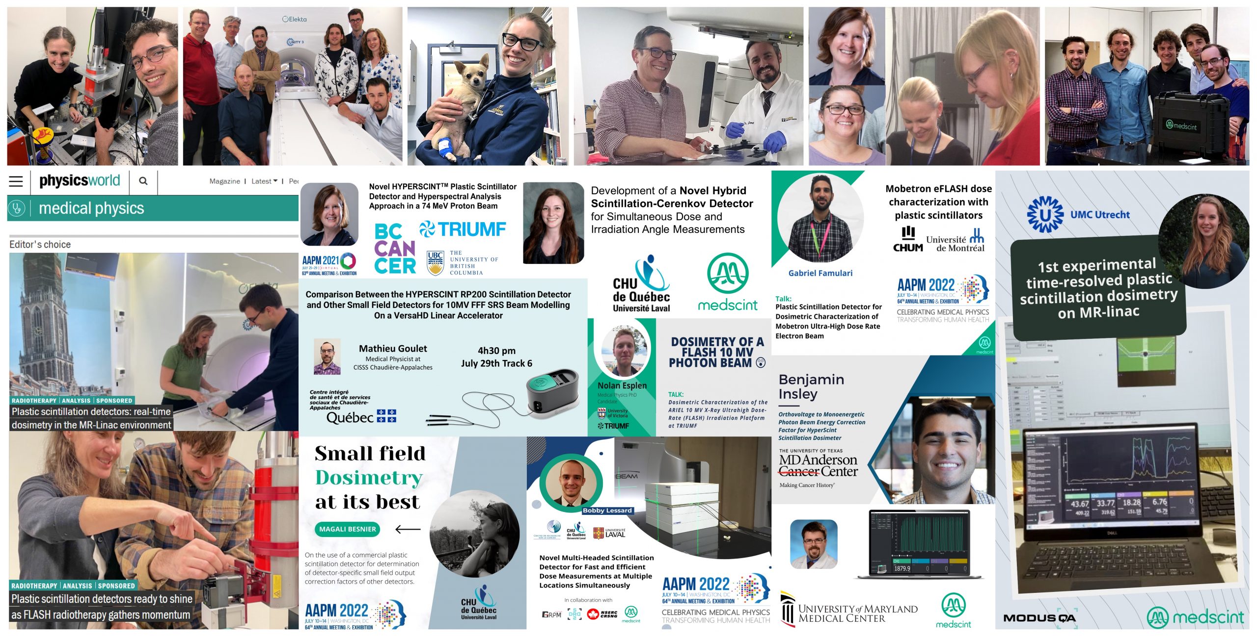
Plastic scintillation detectors: real-time dosimetry in the MR-Linac environment.

Optical innovation meets clinical translation : in the vanguard of adaptive MR/RT research effort, the UMC Utrecht research team works with plastic scintillation detectors to bring MR-Linac treatment to the next level.

PHYSICS WORLD
| UMC Utrecht, Medscint

Plastic scintillation detectors ready to shine as FLASH radiotherapy gathers momentum.

The team of University of Victoria’s XCITE Lab are using plastic scintillation detectors to provide real-time, small-field dosimetry in their FLASH radiotherapy experiments.

PHYSICS WORLD
| University of Victoria – XCITE Lab, Medscint

First Experimental Demonstration of Time-Resolved Plastic Scintillation Dosimetry On An MR-Linac

In this study, UMC Utrecht research team demonstrates the feasibility of a hybrid experimental setup combining an innovative multipoint scintillator detector with film in a moving phantom quantifying MLC tracking for lung SBRT. The prototype cassette is capable of measuring dose (with film and 4 points scintillators simultaneously) during motion experiments, combining film dosimetry with time-resolved and absolute dosimetry.

2022 AAPM ANNUAL MEETING
P.Uijtewaal (1), P.Borman (1), B.Côté (2), Y.Lechasseur (2), J.Turcotte (2), S.Lambert-girard (2), P.Woodhead (1), S.Woodings (1), W.de Vries (1), R.Flores (3), S.Smith (3), B.Raaymakers (1), M.Fast (1) | 1 – UMC Utrecht, Netherland, 2 – Medscint, Canada, 3 – Modus Medical Devices, Canada

Comparison Between the HYPERSCINT RP200 Scintillation Detector and Other Small Field Detectors for 10MV FFF SRS Beam Modelling On a VersaHD Linear Accelerator

Plastic scintillation detectors (PSDs) have advantageous dosimetric properties, including small size and energy independence, which make them ideal candidates for small field dosimetry.

2021 AAPM ANNUAL MEETING
J.Morin, JF.Cabana, M.Goulet, D.Theriault | CISSS – Chaudiere-Appalaches, Lévis, QC, CA

Implementation and validation of beam current transformer for Mobetron ultra-high dose rate electron beam monitoring using multi-detector approach

To evaluate the performance of a custom beam current transformer (BCT) as a beam monitoring tool for the Mobetron electron radiation therapy system at ultra-high dose rates (UHDR) using a multi-detector comparison (plastic scintillators, ion chamber and film).

2022 COMP ANNUAL SCIENTIFIC MEETING
G.Famulari (1), K.Zerouali (1), J.Renaud (2), B.Muir (1), JF.Aubry (1), F.DeBlois (1), JF.Carrier (1) | 1 – Centre Hospitalier de l’Universite de Montreal (CHUM), Montreal, QC, CA, 2 – National Research Council Canada, Montreal, QC, CA

On the orientation independence of the HYPERSCINT scintillation dosimetry research platform in a MR-linac environment.

The purpose of this work was to characterize the HYPERSCINT scintillation dosimetry research platform in a MR-linac environment, particularly with respect to its orientation. This study shows that the HYPERSCINT scintillation dosimetry platform can be used regardless of its orientation in a magnetic field environment. Together with its linearity to dose and dose-rate, the detector shows great promises for development of dosimetry solutions dedicated to the MR-Linac environment.

2022 COMP ANNUAL SCIENTIFIC MEETING
B.Côté (1), B.Raaymakers (2), S.Woodings (2), P.Uijtewaal (2), W.de Vries (2), S.Lambert-girard (1), F.Therriault-Proulx (1), M.Fast (2) | Medscint, Canada (1), UMC Utrecht, Netherland (2)

Pre-clinical and clinical evaluation of the HYPERSCINT plastic scintillation dosimetry research platform for in vivo dosimetry during radiotherapy

The purpose of this work is to evaluate the HYPERSCINT HS-RP100 scintillation dosimetry research platform designed for clinical quality assurance (QA) for use in in vivo dosimetry measurements. The device correctly detected the treatment error when the heads were intentionally laterally shifted. In three canine clinical patients treated in multiple fractions.

J Appl Clin Med Phys. 2022
I.Schoepper (1), S.Dieterich (2), E.Alonzo Trestrail (3), M.Sean Kent (1) | Department of Radiation Oncology, University of California Davis School of Veterinary Medicine, Davis, California, USA, Department of Radiation Oncology, University of California Davis, Medical Center, Sacramento, California, USA, Pacific Crest Medical Physics, Chico, California, USA

Technical note: Characterization and practical applications of a novel plastic scintillator for online dosimetry for an ultrahigh dose rate (FLASH)

Although FLASH radiation therapy is a promising novel technique, the ultrahigh pulsed dose rates mean that experimental dosimetry is very challenging. The plastic scintillator shows a linear and reproducible response and is able to accurately measure the radiation absorbed dose delivered by 16-MeV electrons at UHPDR. The dose is measured accurately in real time with a greater level of precision than that achieved with a radiochromic film.

Med Phys. 2022
Y.Poirier (1,2), J.Xu (1), S.Mossahebi (1), F.Therriault-Proulx (3), A.Sawant (1) | 1- Department of Radiation Oncology, University of Maryland School of Medicine, Maryland, USA, 2- Department of Medical Physics, McGill University, Quebec, Canada, 3- MEDSCINT, Quebec, Canada

Patient Specific QA for External Beam Radiotherapy Using the HYPERSCINT Plastic Scintillation Detector

Plastic scintillation detectors have interesting dosimetric properties, including small size and energy independence. These advantages make them well suited for VMAT patient-specific QA, either alone or in conjunction with a detector matrix. This work aims to determine if the HYPERSCINT scintillation dosimetry research platform can replace the classic ion chamber in a clinical patient-specific QA workflow.

2020 AAPM AM
M.Goulet | CISSS – Chaudiere-Appalaches, Lévis, QC, CA

Characterization of the HYPERSCINT Dosimetry System for Real-Time Dosimetry Measurements with the Varian TrueBeamLinac

Plastic scintillator/optical fibre dosimetry systems are advantageous due to their near water equivalence, waterproof construction, linear dose response, and good spatial resolution due to their small size. The nanosecond decay times of plastic scintillators enable the possibility of real-time dosimetry. We tested the new HYPERSCINT fibre detector system to determine if, in addition to the expected dose and field size responses, this system can provide real-time dose information. The HYPERSCINT system is suitable after appropriate calibration to be used to measure relative dose delivered in cGy as well as indicate changing dose conditions within 0.3 seconds.

2020 AAPM AM
C.Penner (1,2), C.Hoehr (2), C.Mendez (1), C.Duzenli (1) | BC Cancer, Vancouver, BC, CA, TRIUMF, Vancouver, BC, CA

Brachytherapy Technique Commissioning Using the HYPERSCINT Plastic Scintillation Detector

Accurate dosimetry in brachytherapy is not an easy task, as most detectors exhibit volume averaging or energy dependence reducing their usability. Free from these limitations are plastic scintillation detectors, which makes them well suited for brachytherapy applications, either for in vivo dosimetry or commissioning. This work aims to determine if the HYPERSCINT scintillation dosimetry research platform can be used for brachytherapy dose measurement in the context of commissioning a new brachytherapy technique.

2020 AAPM ANNUAL MEETING
M.Goulet, N.Octave, P.Duguay-drouin | CISSS – Chaudiere-Appalaches, Lévis, QC, CA

Characterization of a Novel Plastic Scintillator for Instant Real- Time Dosimetry in Electron FLASH-RT

Purpose was to characterize and validate the novel HYPERSCINT RP100 plastic dosimeter as a direct pulse counter and investigate its use as a real-time in-vivo dosimeter in FLASH-RT radiobiological experiments. In conclusions, the HYPERSCINT RP100 dosimeter accurately measured the delivered radiation absorbed dose under both characterization and biological experimental conditions, with a higher degree of reliability than conventional radiochromic film. Furthermore, its 500 Hz measurement frequency could directly and accurately measure the number of pulses delivered in real time. This shows its potential for real-time in-vivo dosimetry to verify accurate delivery during biological experiments and clinical treatments.

2021 COMP ANNUAL SCIENTIFIC MEETING
Y.Poirier (1), J.Xu (1), A.Ahmady (1), S.Mossahebi (1), H.Zhang (1), F.Therriault-Proulx (2), A.Sawant (1) | 1- University of Maryland School of Medicine, MD, USA , 2- McGill University, QC, Canada, 3- MEDSCINT, QC, Canada

Precise Pulse Delivery Control Using Monitor Units in Electron FLASH-RT

In electron FLASH-RT, precise delivery of the correct number of pulses is critical to accurate dose administration in preclinical radiobiological studies. This work investigates the use of LINAC monitor ion chambers to most precisely control FLASH pulse delivery. Calibrated plastic scintillation detector and EBT-XD Gafchromic films were used for online and passive dosimetry, respectively. The plastic scintillation detector also served as a direct pulse counter.

2021 AAPM ANNUAL MEETING
J.Xu, Y.Poirier, A.Sawant | University of Maryland School of Medicine, MD, USA

Novel Plastic Scintillator for Online Dosimetry in Electron FLASH-RT

The accurate delivery of electrons at FLASH-RT dose rates in radiobiological experiments require new dosimeters that are capable of accurately measuring the radiation dose delivered at >0.55 Gy per pulse (>100 Gy/s) in real-time. The novel HYPERSCINT RP100 plastic dosimeter was able to accurately measure the delivered radiation absorbed dose under characterization and biological experimental conditions, with a higher degree of reliability than conventional radiochromic film. Furthermore, it was shown to directly and accurately measure the number of pulses delivered in real time. This shows potential for use as a real-time in-vivo dosimeter during biological experiments, as well as potential clinical applications.

2021 AAPM ANNUAL MEETING
Y.Poirier (1), J.Xu (1), A.Ahmady (1), S.Mossahebi (1), H.Zhang (1), F.Therriault-Proulx (2), A.Sawant (1) | 1- University of Maryland School of Medicine, Baltimore, MD, USA , 2- MEDSCINT, QC, CANADA

Investigation of the HyperscintTM Plastic Scintillation Dosimetry System Performance in a 15 MeV electron beam at FLASH dose rates

The performance of the HYPERSCINT plastic scintillation dosimetry system in a 15 MeV electron beam operating at FLASH dose rates was investigated. The linac produced a 15 MeV electron FLASH beam with an average dose rate of ~250 Gy/s and dose per pulse between 1 Gy and 1.7 Gy, at the level just above the multi-leaf collimator. The HYPERSCINT plastic scintillator detector system agreed with OSLDs within 3.5% for 10 to 20 pulse FLASH deliveries using the standard dose rate calibration for both dosimeters.

2021 COMP ANNUAL SCIENTIFIC MEETING
C.Mendez, P.Petric, T.Karan, C.Duzenli | BC Cancer, Vancouver, BC, CA

Investigations of a Novel HyperscintTM Plastic Scintillator Detector and Hyperspectral Analysis Approach in a 74 MeV Proton Beam

The HYPERSCINT novel plastic scintillator with spectral analysis approach dosimetry system shows potential for dose measurement in a 74 MeV proton beam with negligible stem effect. The Cerenkov free spectrum may be used to facilitate calibration of the device in MV x-ray beams to improve Cerenkov removal and performance in small field dosimetry.

2021 AAPM ANNUAL MEETING
C.Duzenli (1), C.Hoehr (2), C.Belanger-champagne (2, C.Penner (3), V.Strgar (3) | 1- BC Cancer, BC, CANADA, 2- TRIUMF, BC, CANADA, 3- University of British Columbia, BC, CANADA

EFLASH Dosimetry On a Conventional Linac Using Pulse-Gated Delivery

To build on previous experiments and improve reproducibility of electron FLASH delivery on a conventional linear accelerator, a pulse-gating circuit was constructed and tested with several dosimeters including : a 0.01cc volume ion chamber, optically stimulated luminescence dosimeters (OSLDs), Gafchromic MD film and a novel plastic scintillation detector with spectral analysis (HYPERSCINT).

2021 AAPM ANNUAL MEETING
C.Duzenli, C.Mendez, M.Petric, J.Sweeney, D.Ta, T.Karan | BC Cancer, Vancouver, BC, CANADA

On the nature of the light produced within PMMA optical light guides in scintillation fiber-optic dosimetry

The goal of this study was to evaluate the nature of the stem effect light produced within an optical fiber, to quantify its composition, and to evaluate the efficiency of the chromatic technique to remove the stem effect. The chromatic stem effect removal technique is accurate in most of the situations. However, noticeable differences were obtained between very specific high-energy irradiation conditions. It would be advantageous to implement an additional channel in the chromatic stem effect removal chain or implement a spectral approach to independently remove the Cerenkov and the fluorescence components from the signal of interest. This would increase the accuracy and versatility of the actual chromatic stem effect removal technique.

PHYSICS IN MEDICINE & BIOLOGY
F.Therriault-Proulx (1)(2), L.Beaulieu (2)(3), L.Archambault (2)(3), S.Beddar (4)(1) | 1- Department of Radiation Physics, The University of Texas MD Anderson Cancer Center, Houston, TX, USA, 2- Département de Physique, de Génie Physique et d’Optique, Université Laval, Québec, Québec, Canada, 3- Département de Radio-Oncologie, Hôtel-Dieu de Québec, Centre Hospitalier Universitaire de Québec, Québec, Canada

On the use of a single-fiber multipoint plastic scintillation detector for 192Ir high-dose-rate brachytherapy

The goal of this study was to prove the feasibility of using a single-fiber multipoint plastic scintillation detector as an in vivo verification tool during (192)Ir high-dose-rate brachytherapy treatments. The use of a multipoint plastic scintillation detector for high-dose-rate brachytherapy dosimetry is feasible. This detector shows great promise for development of in vivo applications for real-time verification of treatment delivery.

MEDICAL PHYSICS
F.Therriault-Proulx, S.Beddar, L.Beaulieu | Department of Radiation Physics, The University of Texas MD Anderson Cancer Center, Houston, TX, USA

Development of a novel multi-point plastic scintillation detector with a single optical transmission line for radiation dose measurement

The goal of this study was to develop a novel multi-point plastic scintillation detector capable of measuring the dose accurately at multiple positions simultaneously using a single optical transmission line. This study demonstrates the practical feasibility of multi-point plastic scintillation detector. This type of detector could be very useful for pre-treatment quality assurance applications as well as an accurate tool for real-time in vivo dosimetry.

PHYSICS IN MEDICINE & BIOLOGY
F.Therriault-Proulx, L.Archambault, L.Beaulieu, S.Beddar | Department of Radiation Physics, The University of Texas MD Anderson Cancer Center, Houston, TX, USA, Département de Physique, de Génie Physique et d’Optique, Université Laval, Québec, Québec, Canada, Département de Radio-Oncologie, Hôtel-Dieu de Québec, Centre Hospitalier Universitaire de Québec, Québec, Canada

Review of plastic and liquid scintillation dosimetry for photon, electron, and proton therapy

While scintillation dosimetry has been around for decades, the need for a dosimeter tailored to the reality of modern radiation therapy-in particular a real-time, water-equivalent, energy-independent dosimeter with high spatial resolution-has generated renewed interest in scintillators over the last 10 years. This topical review is intended to provide the medical physics community with a wide overview of scintillation physics, related optical concepts, and applications of plastic scintillation dosimetry.

PHYSICS IN MEDICINE & BIOLOGY
L Beaulieu (1,2), S Beddar (3,4) | 1- Département de physique, génie physique et optique, et Centre de recherche sur le cancer, Université Laval, Québec, CA , 2- Département de radio-oncologie et Axe Oncologie du CRCHU de Québec, QC, CA, 3- Department of Radiation Physics, The University of Texas MD Anderson Cancer Center, Houston, TX, USA, 4- The University of Texas Graduate School of Biomedical Sciences at Houston, Houston, TX, USA

Water-equivalent plastic scintillation detectors for high-energy beam dosimetry: II. Properties and measurements

The properties of a new scintillation detector system for use in dosimetry of high-energy beams in radiotherapy have been measured. The most important properties of these detectors are their hgh spatial resolution and their nearly water-equivalence.

PHYSICS IN MEDICINE & BIOLOGY
A.S. Beddar, T.R. Mackie, F.H. Attix | Depanment of Medical Physics, University of Wisconsin Medical School, Madison, Wl, USA

Water-equivalent plastic scintillation detectors for high-energy beam dosimetry: I. Physical characteristics and theoretical consideration

A minimally perturbing plastic scintillation detector has been developed for the dosimetry of high-energy beams in radiotherapy. The detector system consists of two identical parallel sets of radiation-resistant optical fibre bundles, each connected to independent photomultiplier tubes.

PHYSICS IN MEDICINE & BIOLOGY
A.S. Beddar, T.R. Mackie, F.H. Attix | Depanment of Medical Physics, University of Wisconsin Medical School, Madison, Wl, USA

A method to correct for temperature dependence and measure simultaneously dose and temperature using a plastic scintillation detector

Plastic scintillation detectors work well for radiation dosimetry. However, they show some temperature dependence, and a priori knowledge of the temperature surrounding the plastic scintillation detectors is required to correct for this dependence.

PHYSICS IN MEDICINE & BIOLOGY
F.Therriault-Proulx, L.Wooton, S.Beddar | Department of Radiation Physics, The University of Texas MD Anderson Cancer Center, Houston, TX, USA

Beam matching for small-field dosimetry applications using accelerator solenoid current and a miniature plastic scintillation detector.

The goal of this work is to determine the optimal accelerator solenoid current (ASOL) that minimizes the spread of measured small field output factor (OF) values between a series of machines. Small field OF measurements of 6 MV flattening filtered beams from 3 Varian TrueBeam linear accelerators were performed using a 1 mm diameter by 1 mm length plastic scintillation detector HYPERSCINT HS-RP200 research platform. In conclusion Small field dosimetry characteristics are highly sensitive to beam focal spot size. and the possibility to optimize accelerator focusing coil current to reduce OF and penumbra width spread enables new avenues in beam matching of series of machines, especially for SRS and SBRT techniques.

2022 COMP ANNUAL SCIENTIFIC MEETING
L.Gingras (1), F.Beaulieu (1), M.Besnier (1), B.Côté (2), D.Leblanc (2), L.Beaulieu (1), L.Archambault (1) | 1- CHU de Québec – Université Laval, QC, CA, 2- MEDSCINT, QC, CA

FLASH Irradiation of Drosophila Melanogaster Using Low Energy X-Rays

To investigate the capability of low energy x-rays to elicit the FLASH effect, Drosophila melanogaster larvae were irradiated with ultrahigh dose-rate and conventional radiotherapy dose rates using an inexpensive x-ray tube system. Dosimetry was performed with plastic scintillators and radiochromic film, and the differential survival observed in this work suggests that continuous 120 kVp x-rays can induce a FLASH effect.

2022 AAPM ANNUAL MEETING
A.Hart, J.Dudzic, J.Eby, S.Perlman, M.Bazalova-Carter | University of Victoria, Victoria, BC ,CA

Orthovoltage to Monoenergetic Photon Beam Energy Correction Factor for HyperScint Scintillation Dosimeter

The scintillator dosimetry system is a small-field dosimeter with reported energy independence down to 100-keV. This work investigates the energy dependence of the scintillator between a monoenergetic photon source and polyenergetic orthovoltage beam.

2022 AAPM ANNUAL MEETING
B. Insley, D. Bartkoski, L. Che Fru, M. Salehpour | M.D. Anderson Cancer Center, Houston, TX

Dosimetric Characterization of the ARIEL 10 MV X-Ray Ultrahigh Dose-Rate (FLASH) Irradiation Platform at TRIUMF

The purpose was to characterize the beam delivery capabilities and dose rates achievable on the new ultrahigh dose-rate 10MV x-ray irradiation platform at TRIUMF. Beam commissioning and dosimetry have been conducted on the ARIEL x-ray FLASH irradiation platform using film doses and scintillators. Measured dose rates support that the 10MV x-ray beam may be used as a UHDR source compatible with FLASH radiobiological experiments.

2022 AAPM ANNUAL MEETING
N.Esplen (1), L.Egoriti (2), T.Planche (3), A.Hart (1), B.Paley (3), C.Hoehr (3), A.Gottberg (3), M.Bazalova-Carter (1) | 1- University of Victoria, BC ,CA, 2- University of British Columbia, BC, CA, 3- TRIUMF, BC, CA

Plastic Scintillation Detector for Dosimetric Characterization of Mobetron Ultra-High Dose Rate Electron Beam

The purpose is to present commissioning data for the MOBETRON electron radiation therapy system (IntraOp) at ultra-high dose rate using the HYPERSCINT plastic scintillation detector. The suitability of using a plastic scintillator as an active dosimeter for commissioning measurements of an ultra-high dose rate electron beam has been demonstrated (reference dosimetry, DPP, beam penetration, linearity with number of pulses, linearity with PW and short-term output stability).

2022 AAPM ANNUAL MEETING
G.Famulari (1), K.Zerouali (1), O.Piron (1), JF.Aubry (1), F.DeBlois (1), JF.Carrier (2) | 1- Centre Hospitalier de l’Universite de Montreal (CHUM), Montreal, QC, CA, 2- Departement de Physique, Universite de Montreal, Montreal, QC, CA

A Novel Multi-Headed Scintillation Detector for Fast and Efficient Dose Measurements at Multiple Locations Simultaneously

To develop and quantify the performances of a novel multi-point scintillation detector having multiple heads connected to the same optical line, allowing real-time dose measurements simultaneously at 3 positions in non-contiguous space.

2022 AAPM ANNUAL MEETING
B.Lessard (1,2), Y.Lechasseur (3), S.Lambert-girard (3), F.Therriault-Proulx (3), L.Beaulieu (1,2), L.Archambault (1,2) | 1- Département de physique, génie physique et optique, et Centre de recherche sur le cancer, Université Laval, Québec, CA , 2- CHU de Quebec – Universite Laval, QC, Canada, 3- MEDSCINT, QC, Canada

Quantifying the DNA-damaging Effects of FLASH Irradiation With Plasmid DNA

The objective is to investigate a plasmid DNA nicking assay approach for isolating and quantifying the DNA-damaging effects of ultrahigh-dose-rate (ie FLASH) irradiation relative to conventional dose-rate irradiation. The doses and dose rates were verified independently using EBT-XD Gafchromic film placed directly above the DNA-based phantom and HYPERSCINT high temporal resolution plastic scintillator placed immediately beside the DNA phantoms (both phantoms had been previously calibrated at conventional dose rates and validated at FLASH-RT dose rates).

INTERNATIONAL JOURNAL OF RADIATION ONCOLOGY, BIOLOGY, PHYSICS
A.Perstin (1), Y.Poirier (2), A.Sawant (2), M.Tambasco (1) | 1- Department of Physics, San Diego State University, CA, USA, 2- Department of Radiation Oncology, University of Maryland School of Medicine, Maryland, USA

External beam irradiation angle measurement using Cerenkov emission

In this study, we propose a novel approach designed to take advantage of the Cerenkov angular dependency to perform a direct measurement of an external beam radiation angle of incidence. The detector offers promising perspectives for external beam radiotherapy and brachytherapy applications.

MEDICAL PHYSICS
E.Jean (1,2,3), S.Lambert-girard (4), F.Therriault-Proulx (4), L.Beaulieu (1,2) | 1- Département de physique, génie physique et optique, et Centre de recherche sur le cancer, Université Laval, QC, Canada, 2- Département de radio-oncologie et Axe Oncologie du CRCHU de Québec, QC, CA, 3- Département de radio-oncologie du CIUSSS-MCQ, CHAUR de Trois-Rivières, QC, Canada, 4- MEDSCINT, QC, Canada

Characterization of an x‐ray tube‐based ultrahigh dose‐rate system for in vitro irradiations.

To present an x-ray tube system capable of in vitro ultrahigh dose-rate (UHDR) irradiation of small < 0.3 mm samples and to characterize it by means of a plastic scintillation detector (PSD).

Med Phys. 2021
D.Cecchi (1), F.Therriault-Proulx (2), S.Lambert-girard (2), A.Hart (1), A.Macdonald (1), M.Pfleger (1), M.Lenckowski (1), M.Bazalova-Carter (1) | Department of Physics and Astronomy, University of Victoria, Victoria, British Columbia, Canada, MedScint, QC, CA