High-throughput, low-cost FLASH: irradiation of Drosophila melanogaster with low-Energy X-rays using time structures spanning ConvDR and UHDR

This article explores the potential of using low-energy X-rays to deliver ultrahigh dose-rate (UHDR) FLASH radiotherapy using Drosophila melanogaster as a model. For this they have compared the effects of UHDR (210 Gy/s) and conventional dose rates (0.2–0.4 Gy/s) on the eclosion and lifespan of fly larvae. The results showed that larvae treated with UHDR had higher survival rates and longer lifespans, particularly at intermediate doses, indicating a normal tissue-sparing FLASH effect.

The Medscint scintillation dosimetry detector was used to measure the response to X-rays at a very high sampling rate to confirm the time structure of the delivered radiation (i.e. the pulse width and inter-pulse spacing). Along with film measurements, they also confirmed that the doses delivered with UHDR and CONV agreed within 0.1%.
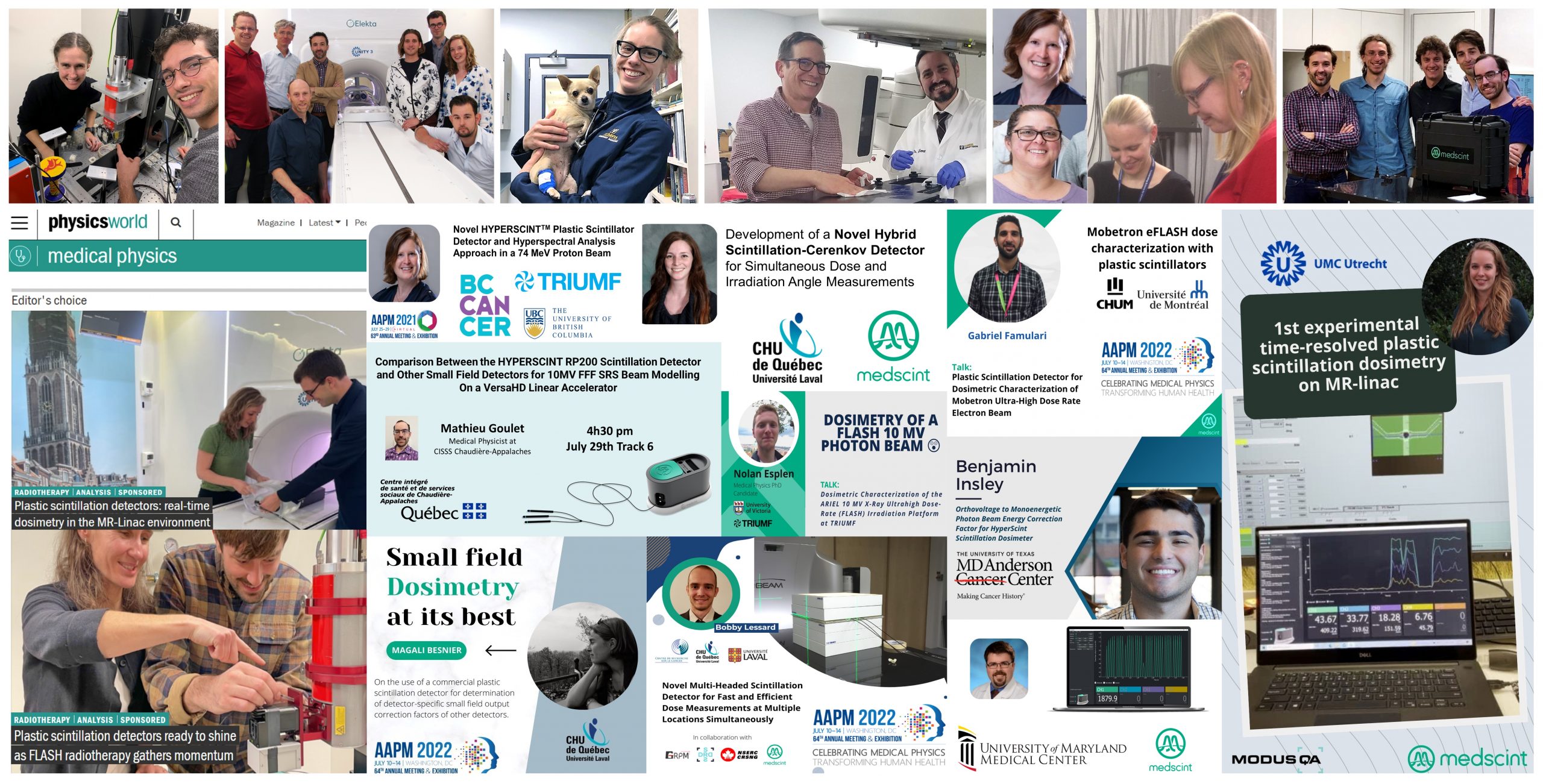
Journal of Radiation Research
Alexander Hart (1), Jan P Dudzic (1), Jameson W Clarke (1), Jonathan Eby (2), Steve J Perlman (1), Magdalena Bazalova-Carter (1) | 1. University of Victoria, BC – CANADA, 2. University of Toronto, ON – CANADA

Development of End-to-End Preclinical Treatment Verification Procedures, Traceable to NPL Air Kerma Primary Standard

Dosimetry audits are an important tool to improve quality of reported results and to support standardization of preclinical radiation research. This work presents how the combination of passive and active detectors, such as the real-time HYPERSCINT scintillation dosimetry solution, with anatomically correct mouse phantoms are adequate for the development of End-to-End dosimetry audits for the independent verification of preclinical radiation treatments.

The traceability of the detectors’ calibration to primary standards strengthens the dosimetry chain in the validation of preclinical plans, and it is consistent with the current practice for dose traceability of clinical radiotherapy treatments. Their implementation at national and regional levels could lead to databases of anonymised records, which will positively impact the dissemination of best practices and sharing of validated results.

6th Conference on small animal precision image-guided radiotherapy
Ileana Silvestre Patallo (1), Rebecca Carter (2)(3), Andrew Nisbet (2), Anna Subiel (1), Giuseppe Schettino (1) | 1. National Physical Laboratory, UK, 2. University College London, UK, 3. Cancer Institut, UK

Technical note: Characterization and practical applications of a novel plastic scintillator for online dosimetry for an ultrahigh dose rate (FLASH)

Although FLASH radiation therapy is a promising novel technique, the ultrahigh pulsed dose rates mean that experimental dosimetry is very challenging. The plastic scintillator shows a linear and reproducible response and is able to accurately measure the radiation absorbed dose delivered by 16-MeV electrons at UHPDR. The dose is measured accurately in real time with a greater level of precision than that achieved with a radiochromic film.

Med Phys. 2022
Y.Poirier (1,2), J.Xu (1), S.Mossahebi (1), F.Therriault-Proulx (3), A.Sawant (1) | 1- Department of Radiation Oncology, University of Maryland School of Medicine, Maryland, USA, 2- Department of Medical Physics, McGill University, Quebec, Canada, 3- MEDSCINT, Quebec, Canada

Novel Plastic Scintillator for Online Dosimetry in Electron FLASH-RT

The accurate delivery of electrons at FLASH-RT dose rates in radiobiological experiments require new dosimeters that are capable of accurately measuring the radiation dose delivered at >0.55 Gy per pulse (>100 Gy/s) in real-time. The novel HYPERSCINT RP100 plastic dosimeter was able to accurately measure the delivered radiation absorbed dose under characterization and biological experimental conditions, with a higher degree of reliability than conventional radiochromic film. Furthermore, it was shown to directly and accurately measure the number of pulses delivered in real time. This shows potential for use as a real-time in-vivo dosimeter during biological experiments, as well as potential clinical applications.

2021 AAPM ANNUAL MEETING
Y.Poirier (1), J.Xu (1), A.Ahmady (1), S.Mossahebi (1), H.Zhang (1), F.Therriault-Proulx (2), A.Sawant (1) | 1- University of Maryland School of Medicine, Baltimore, MD, USA , 2- MEDSCINT, QC, CANADA

FLASH Irradiation of Drosophila Melanogaster Using Low Energy X-Rays

To investigate the capability of low energy x-rays to elicit the FLASH effect, Drosophila melanogaster larvae were irradiated with ultrahigh dose-rate and conventional radiotherapy dose rates using an inexpensive x-ray tube system. Dosimetry was performed with plastic scintillators and radiochromic film, and the differential survival observed in this work suggests that continuous 120 kVp x-rays can induce a FLASH effect.

2022 AAPM ANNUAL MEETING
A.Hart, J.Dudzic, J.Eby, S.Perlman, M.Bazalova-Carter | University of Victoria, Victoria, BC ,CA

Quantifying the DNA-damaging Effects of FLASH Irradiation With Plasmid DNA

The objective is to investigate a plasmid DNA nicking assay approach for isolating and quantifying the DNA-damaging effects of ultrahigh-dose-rate (ie FLASH) irradiation relative to conventional dose-rate irradiation. The doses and dose rates were verified independently using EBT-XD Gafchromic film placed directly above the DNA-based phantom and HYPERSCINT high temporal resolution plastic scintillator placed immediately beside the DNA phantoms (both phantoms had been previously calibrated at conventional dose rates and validated at FLASH-RT dose rates).

INTERNATIONAL JOURNAL OF RADIATION ONCOLOGY, BIOLOGY, PHYSICS
A.Perstin (1), Y.Poirier (2), A.Sawant (2), M.Tambasco (1) | 1- Department of Physics, San Diego State University, CA, USA, 2- Department of Radiation Oncology, University of Maryland School of Medicine, Maryland, USA

Characterization of an x‐ray tube‐based ultrahigh dose‐rate system for in vitro irradiations.

To present an x-ray tube system capable of in vitro ultrahigh dose-rate (UHDR) irradiation of small < 0.3 mm samples and to characterize it by means of a plastic scintillation detector (PSD).

Med Phys. 2021
D.Cecchi (1), F.Therriault-Proulx (2), S.Lambert-girard (2), A.Hart (1), A.Macdonald (1), M.Pfleger (1), M.Lenckowski (1), M.Bazalova-Carter (1) | Department of Physics and Astronomy, University of Victoria, Victoria, British Columbia, Canada, MedScint, QC, CA