Development of End-to-End Preclinical Treatment Verification Procedures, Traceable to NPL Air Kerma Primary Standard

Dosimetry audits are an important tool to improve quality of reported results and to support standardization of preclinical radiation research. This work presents how the combination of passive and active detectors, such as the real-time HYPERSCINT scintillation dosimetry solution, with anatomically correct mouse phantoms are adequate for the development of End-to-End dosimetry audits for the independent verification of preclinical radiation treatments.

The traceability of the detectors’ calibration to primary standards strengthens the dosimetry chain in the validation of preclinical plans, and it is consistent with the current practice for dose traceability of clinical radiotherapy treatments. Their implementation at national and regional levels could lead to databases of anonymised records, which will positively impact the dissemination of best practices and sharing of validated results.
6th Conference on small animal precision image-guided radiotherapy
Ileana Silvestre Patallo (1), Rebecca Carter (2)(3), Andrew Nisbet (2), Anna Subiel (1), Giuseppe Schettino (1) | 1. National Physical Laboratory, UK, 2. University College London, UK, 3. Cancer Institut, UK

Plastic scintillator dosimetry of ultrahigh dose-rate 200 MeV electrons at CLEAR

Very high energy electron (VHEE) beams with energies greater than 100 MeV may be promising candidates for FLASH radiotherapy due to their favourable dose distributions and accessibility of ultrahigh dose-rates (UHDR). The standard dosimeters used for conventional radiotherapy, including ionization chambers and film, have limited application to UHDR radiotherapy due to deficits in dose rate independence and temporal resolution. The performance of PSDs in this work suggest they may be useful real-time dosimeters for applications in UHDR VHEE radiotherapy.

IEEE Xplore
Alexander Hart (1), Cloé Giguère (2,6), Joseph Bateman (3,4), Pierre Korysko (3,4), Wilfrid Farabolini (3), Vilde Rieker (3,5), Nolan Esplen (1), Roberto Corsini (3), Manjit Dosanjh (3,4), Luc Beaulieu(2,6), Magdalena Bazalova-Carter (1) | 1. Department of Physics and Astronomy, University of Victoria, Victoria, BC, Canada, 2. Département de Physique, de génie Physique et d’optique et Centre de Recherche sur le Cancer, Université Laval, Quebec, QC, Canada, 3. CERN, Geneva, Switzerland, 4. Department of Physics, University of Oxford, Oxford, United Kingdom, 5. Department of Physics, University of Oslo, Oslo, Norway, 6. Département de radio-oncologie et Axe Oncologie duCRCHUde Québec, CHUde Québec – Universit é Laval, Quebec, QC, Canada

Investigation of temperature dependence of inorganic scintillators using the HYPERSCINT research platform

The temperature dependence of four inorganic scintillation detectors was examined spectrally using the HYPERSCINT Research Platform 200 under 6 MV photon irradiations from a LINAC. After varying only the temperature of the detectors, all scintillators demonstrated linearity when the change in photon counts with temperature in the full-width at half maximum of their spectrum are integrated. Establishing the magnitude of the temperature dependence of the materials is critical to decide whether correction factors are required. This is especially true in applications such as brachytherapy, where detectors equilibrise to body temperature.

Radiation Measurements
Owen McLaughlin (1), Michael Martyn (1,2), Christoph Kleefeld (1), Mark Foley (1) | 1. Physics Unit, School of Natural Sciences, University of Galway, Galway, Ireland, 2. Galway Clinic, Doughiska, Galway, Ireland

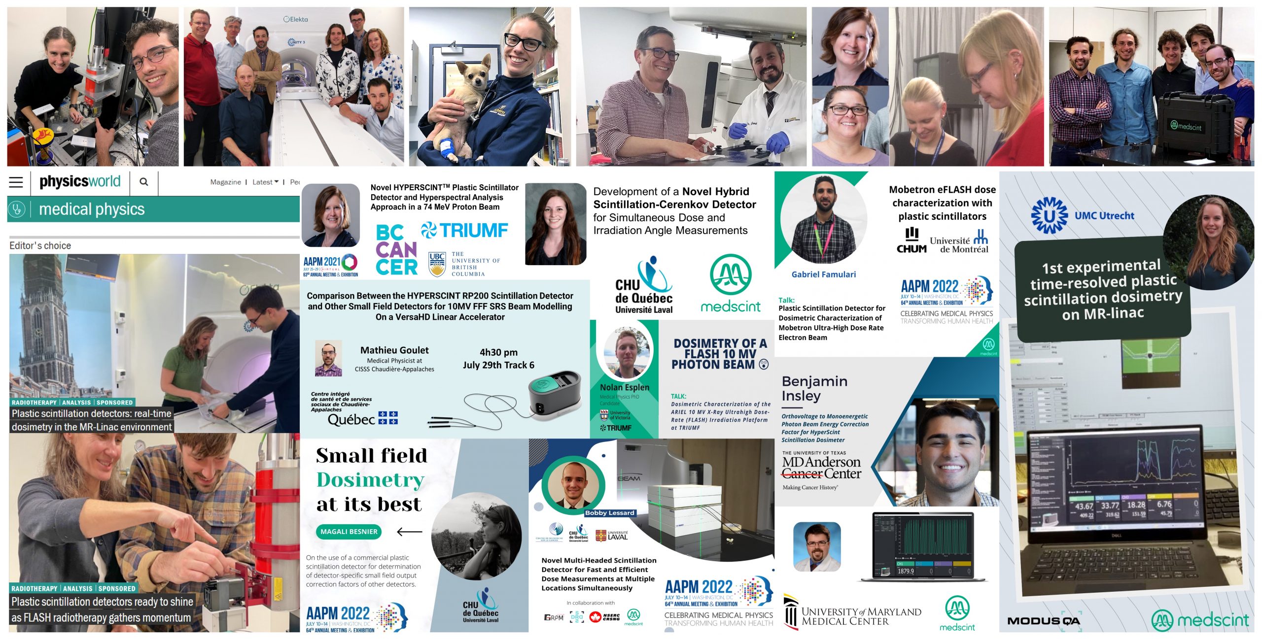
Plastic scintillation detectors: real-time dosimetry in the MR-Linac environment.

Optical innovation meets clinical translation : in the vanguard of adaptive MR/RT research effort, the UMC Utrecht research team works with plastic scintillation detectors to bring MR-Linac treatment to the next level.

PHYSICS WORLD
| UMC Utrecht, Medscint

Plastic scintillation detectors ready to shine as FLASH radiotherapy gathers momentum.

The team of University of Victoria’s XCITE Lab are using plastic scintillation detectors to provide real-time, small-field dosimetry in their FLASH radiotherapy experiments.

PHYSICS WORLD
| University of Victoria – XCITE Lab, Medscint

Investigations of a Novel HyperscintTM Plastic Scintillator Detector and Hyperspectral Analysis Approach in a 74 MeV Proton Beam

The HYPERSCINT novel plastic scintillator with spectral analysis approach dosimetry system shows potential for dose measurement in a 74 MeV proton beam with negligible stem effect. The Cerenkov free spectrum may be used to facilitate calibration of the device in MV x-ray beams to improve Cerenkov removal and performance in small field dosimetry.

2021 AAPM ANNUAL MEETING
C.Duzenli (1), C.Hoehr (2), C.Belanger-champagne (2, C.Penner (3), V.Strgar (3) | 1- BC Cancer, BC, CANADA, 2- TRIUMF, BC, CANADA, 3- University of British Columbia, BC, CANADA

EFLASH Dosimetry On a Conventional Linac Using Pulse-Gated Delivery

To build on previous experiments and improve reproducibility of electron FLASH delivery on a conventional linear accelerator, a pulse-gating circuit was constructed and tested with several dosimeters including : a 0.01cc volume ion chamber, optically stimulated luminescence dosimeters (OSLDs), Gafchromic MD film and a novel plastic scintillation detector with spectral analysis (HYPERSCINT).

2021 AAPM ANNUAL MEETING
C.Duzenli, C.Mendez, M.Petric, J.Sweeney, D.Ta, T.Karan | BC Cancer, Vancouver, BC, CANADA

On the nature of the light produced within PMMA optical light guides in scintillation fiber-optic dosimetry

The goal of this study was to evaluate the nature of the stem effect light produced within an optical fiber, to quantify its composition, and to evaluate the efficiency of the chromatic technique to remove the stem effect. The chromatic stem effect removal technique is accurate in most of the situations. However, noticeable differences were obtained between very specific high-energy irradiation conditions. It would be advantageous to implement an additional channel in the chromatic stem effect removal chain or implement a spectral approach to independently remove the Cerenkov and the fluorescence components from the signal of interest. This would increase the accuracy and versatility of the actual chromatic stem effect removal technique.

PHYSICS IN MEDICINE & BIOLOGY
F.Therriault-Proulx (1)(2), L.Beaulieu (2)(3), L.Archambault (2)(3), S.Beddar (4)(1) | 1- Department of Radiation Physics, The University of Texas MD Anderson Cancer Center, Houston, TX, USA, 2- Département de Physique, de Génie Physique et d’Optique, Université Laval, Québec, Québec, Canada, 3- Département de Radio-Oncologie, Hôtel-Dieu de Québec, Centre Hospitalier Universitaire de Québec, Québec, Canada

On the use of a single-fiber multipoint plastic scintillation detector for 192Ir high-dose-rate brachytherapy

The goal of this study was to prove the feasibility of using a single-fiber multipoint plastic scintillation detector as an in vivo verification tool during (192)Ir high-dose-rate brachytherapy treatments. The use of a multipoint plastic scintillation detector for high-dose-rate brachytherapy dosimetry is feasible. This detector shows great promise for development of in vivo applications for real-time verification of treatment delivery.

MEDICAL PHYSICS
F.Therriault-Proulx, S.Beddar, L.Beaulieu | Department of Radiation Physics, The University of Texas MD Anderson Cancer Center, Houston, TX, USA

Development of a novel multi-point plastic scintillation detector with a single optical transmission line for radiation dose measurement

The goal of this study was to develop a novel multi-point plastic scintillation detector capable of measuring the dose accurately at multiple positions simultaneously using a single optical transmission line. This study demonstrates the practical feasibility of multi-point plastic scintillation detector. This type of detector could be very useful for pre-treatment quality assurance applications as well as an accurate tool for real-time in vivo dosimetry.

PHYSICS IN MEDICINE & BIOLOGY
F.Therriault-Proulx, L.Archambault, L.Beaulieu, S.Beddar | Department of Radiation Physics, The University of Texas MD Anderson Cancer Center, Houston, TX, USA, Département de Physique, de Génie Physique et d’Optique, Université Laval, Québec, Québec, Canada, Département de Radio-Oncologie, Hôtel-Dieu de Québec, Centre Hospitalier Universitaire de Québec, Québec, Canada