Quantifying the DNA-damaging Effects of FLASH Irradiation With Plasmid DNA

The objective is to investigate a plasmid DNA nicking assay approach for isolating and quantifying the DNA-damaging effects of ultrahigh-dose-rate (ie FLASH) irradiation relative to conventional dose-rate irradiation. The doses and dose rates were verified independently using EBT-XD Gafchromic film placed directly above the DNA-based phantom and HYPERSCINT high temporal resolution plastic scintillator placed immediately beside the DNA phantoms (both phantoms had been previously calibrated at conventional dose rates and validated at FLASH-RT dose rates).

INTERNATIONAL JOURNAL OF RADIATION ONCOLOGY, BIOLOGY, PHYSICS
A.Perstin (1), Y.Poirier (2), A.Sawant (2), M.Tambasco (1) | 1- Department of Physics, San Diego State University, CA, USA, 2- Department of Radiation Oncology, University of Maryland School of Medicine, Maryland, USA

External beam irradiation angle measurement using Cerenkov emission

In this study, we propose a novel approach designed to take advantage of the Cerenkov angular dependency to perform a direct measurement of an external beam radiation angle of incidence. The detector offers promising perspectives for external beam radiotherapy and brachytherapy applications.

MEDICAL PHYSICS
E.Jean (1,2,3), S.Lambert-girard (4), F.Therriault-Proulx (4), L.Beaulieu (1,2) | 1- Département de physique, génie physique et optique, et Centre de recherche sur le cancer, Université Laval, QC, Canada, 2- Département de radio-oncologie et Axe Oncologie du CRCHU de Québec, QC, CA, 3- Département de radio-oncologie du CIUSSS-MCQ, CHAUR de Trois-Rivières, QC, Canada, 4- MEDSCINT, QC, Canada

Characterization of an x‐ray tube‐based ultrahigh dose‐rate system for in vitro irradiations.

To present an x-ray tube system capable of in vitro ultrahigh dose-rate (UHDR) irradiation of small < 0.3 mm samples and to characterize it by means of a plastic scintillation detector (PSD).

Med Phys. 2021
D.Cecchi (1), F.Therriault-Proulx (2), S.Lambert-girard (2), A.Hart (1), A.Macdonald (1), M.Pfleger (1), M.Lenckowski (1), M.Bazalova-Carter (1) | Department of Physics and Astronomy, University of Victoria, Victoria, British Columbia, Canada, MedScint, QC, CA

Comparative optic and dosimetric characterization of the HYPERSCINT scintillation dosimetry research platform for multipoint applications

This study introduces the HYPERSCINT research platform (HYPERSCINT-RP100, Medscint Inc., Quebec, Canada), the first commercially available scintillation dosimetry platform capable of multi-point dosimetry through the hyperspectral approach.

Phys Med Biol. 2021
E.Jean (1,2,3), F.Therriault-Proulx (4), L.Beaulieu (1,2) | Département de physique, génie physique et optique, et Centre de recherche sur le cancer, Université Laval, Québec, CA , Département de radio-oncologie et Axe Oncologie du CRCHU de Québec, QC, CA, Département de radio-oncologie du CIUSSS-MCQ, CHAUR de Trois-Rivières, Trois-Rivières, QC, Canada, MedScint, QC, CA

Pre-Clinical and Clinical Evaluation of the HYPERSCINT Scintillation Dosimetry Research Platform

The purpose of this work is to evaluate the HYPERSCINT scintillation dosimetry research platform (Medscint Inc., Quebec City, Canada) designed for clinical QA for use in in-vivo dosimetry measurements.

2020 AAPM AM
I.Schoepper, E.Trestrail, S.Dieterich, M.Kent | WR Pritchard Veterinary Medical Teaching Hospital, UC Davis School of Veterinary Medicine UC Davis School Of Veterinary Medicine, Pacific Crest Medical Physics ,Chico, CA, UC Davis Medical Center, Sacramento, CA, Surgical and Radiological Sciences, UC Davis School of Veterinary Medicine, Davis, CA

Dosimetric Characterization of the HYPERSCINT Scintillation Dosimetry Research Platform for Multipoint Applications

Multipoint detectors are very interesting toward developing new phantoms and making measurements in constrained space. This study aims at assessing the performance of the HYPERSCINT dosimetry research platform (Medscint inc., Québec, Canada) with a high spatial resolution 3-point plastic scintillation detector for application to high energy photon beam dosimetry.

2020 AAPM AM
E.Jean, F.Therriault-Proulx, L.Beaulieu | CHU de Quebec – Universite Laval, QC, CA, Medscint, QC, CA , Centre Hospitalier Univ de Québec, Québec, QC, CA

Development of An Automated Routine for Finding the Precise Location of Scintillators Elements and Their Emission Spectrum in a Multi-Point Scintillation Detector

The goal of this study is to develop an approach allowing for calibration of multi-point scintillation detector (mPSD) using only the photon beam from a linear accelerator such that it doesn’t depend on the availability of other irradiation modalities (e.g. orthovoltage irradiators). This study also aims at determining an experimental method to validate the spatial position of the different scintillators within the mPSD.

2020 AAPM AM
B.Lessard (1,2), F.Larose (1,2), F.Berthiaume (1,2,3), S.Lambert-girard (1,2,3), F.Therriault-Proulx (3), L.Archambault (1,2) | Département de physique, génie physique et optique, et Centre de recherche sur le cancer, Université Laval, Québec, CA , CHU de Quebec – Universite Laval, QC, CA, MedScint, QC, CA

Feasibility of Plastic Scintillator Dosimeters for FLASH Therapy

To examine the capabilities of plastic scintillator dosimeters (PSDs) to accurately measure FLASH radiotherapy dose rates delivered with an x-ray tube.

2020 AAPM AM
D.Cecchi (1), F.Therriault-Proulx (2), M.Bazalova-Carter (1) | University of Victoria, Victoria, BC ,CA,, Medscint, QC, CA

Development and characterization of an optical fiber-based Cerenkov dosimeter

This study introduces a novel hybrid Cerenkov-scintillation dosimeter

2021 COMP ASM
E.Jean (1,2,3), S.Lambert-girard (3), F.Therriault-Proulx (3), L.Beaulieu (1,2) | CHU de Quebec – Universite Laval, QC, CA, Département de radio-oncologie et Axe Oncologie du CRCHU de Québec, QC, CA, MedScint, QC, CA
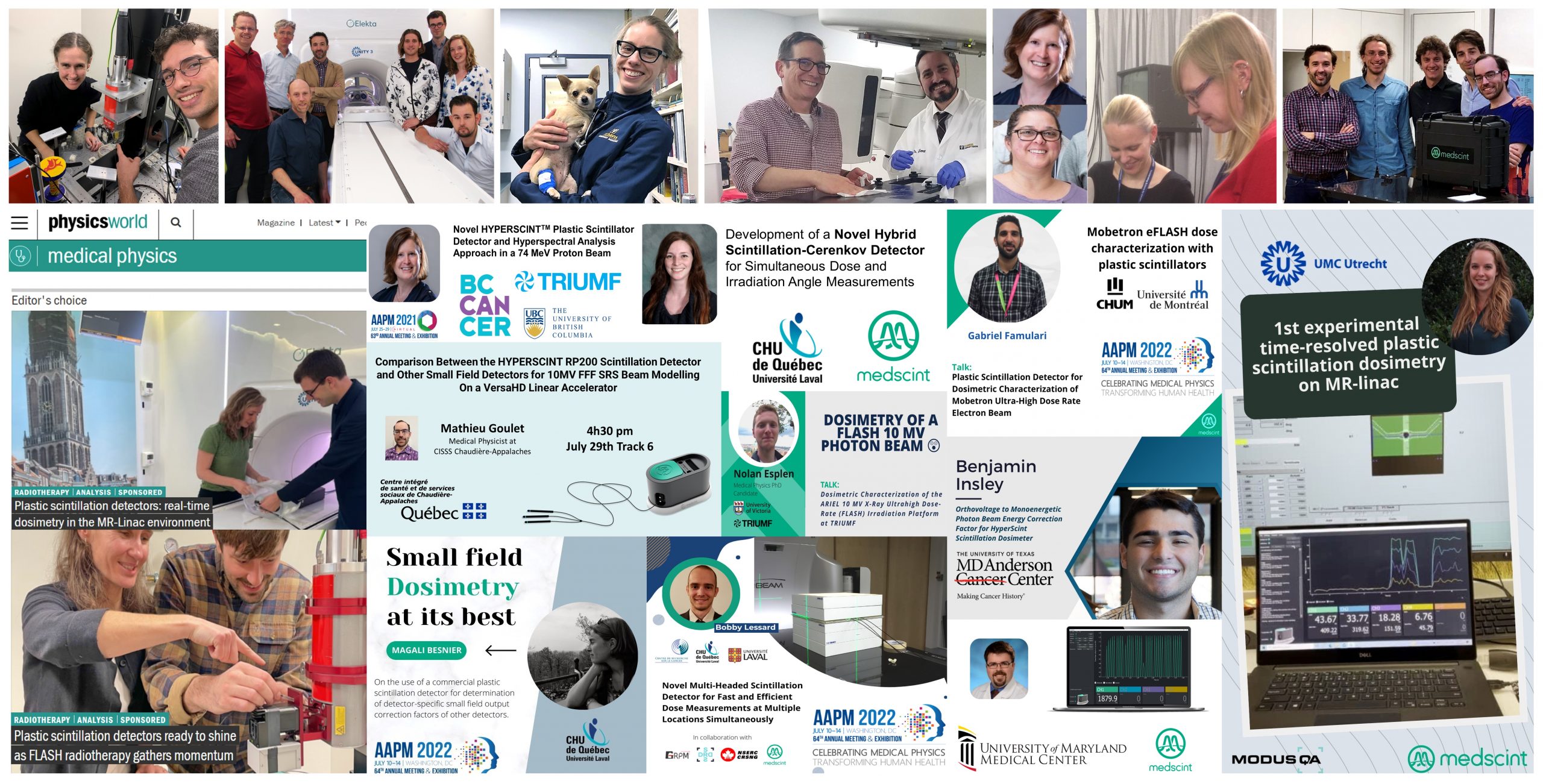
Small field dosimetry of a Varian TrueBeam High Definition MLC linear accelerator using theHyperscint RP200 scintillation detector.

Small field dosimetry of a Varian TrueBeam High Definition MLC linear accelerator using the Hyperscint RP200 scintillation detector.

Purpose: To evaluate the performance of the new Hyperscint RP200 plastic scintillator for small field measurements of a Varian TrueBeam linear accelerator in comparison with the current state-of-the-art methodology in the clinic.

Methods: Small field measurements were performed using different detectors: a diamond detector (microDiamond, PTW), a diode (Razor, IBA), a compact ion chamber (Razor, IBA), and a 1mm x 1mm
plastic scintillation detector (Hyperscint RP200, Medscint). Correction factors based on measured field sizes, following TRS483 recommendations, were applied to all measurements. Output factors of a
TrueBeam linear accelerator were measured for field sizes of 0.5 cm to 2 cm for jaws and MLC configurations for 6-MV, 6-MV FFF and 10-MV FFF photon beams. Output factors for different circular collimators (0.4 cm to 2 cm) were also obtained at 10-MV FFF. Scintillator measurements were compared to the small-field dosimetry methodology used clinically.

Results: No correction factors were necessary for the plastic scintillation detector measurements. Scintillator measurements were within 1.1% of the standard methodology for all the small field geometries studied. Average relative differences were (0.3±0.5)%, (0.7±0.3)%, and (0.2±0.2)% for the 6-MV, 6-MV FFF, and 10-MV FFF, respectively. Output factors of circular field sizes down to 0.4-cm diameter were obtained with an average relative difference of (0.1±0.4)%, including a maximum difference of 0.7% for the smallest field.

Conclusion: This new scintillation dosimetry research platform shows great promises for small field dosimetry. It has the potential to be used as part of a single-detector no-correction-factor methodology.

2021 COMP ASM
L.Gingras, B.Côté, F.Berthiaume, S.Lambert-Girard, D.Leblanc, L.Archambault, L.Beaulieu, F.Therriault-Proulx | CHU de Quebec – Universite Laval, QC, CA, Département de radio-oncologie et Axe Oncologie du CRCHU de Québec, QC, CA, MedScint, QC, CA

Development of a Novel Hybrid Scintillation-Cerenkov Detector for Simultaneous Dose and Irradiation Angle Measurements

This study introduces a novel hybrid detector capable of simultaneous dose and direct irradiation angle measurements based on Cerenkov angular dependency.

2021 AAPM AM
E.Jean (1,2), S.Lambert-girard (3), F.Therriault-Proulx (3), L.Beaulieu (1) | CHU de Quebec – Universite Laval, QC, CA, CHAUR, Trois-Rivieres, QC, CA , MedScint, QC, CA

Plastic and Lead-Doped Scintillators for Ultrahigh Dose-Rate Irradiations Delivered with An X-Ray Tube

To examine the capabilities of plastic scintillators of different compositions to accurately measure dose in high dose-rate dose irradiations delivered with an x-ray tube.

2021 AAPM AM
D.Cecchi (1), C.Gigeure (2), F.Larose (2), F.Therriault-Proulx (3), L.Beaulieu (2), M.Bazalova-Carter (1) | University of Victoria, Victoria, BC ,CA,, CHU de Quebec – Universite Laval, QC, CA, MedScint, QC, CA

Evaluation of scintillation detectors for ultrahigh dose-rate x-ray beam dosimetry

FLASH-Radiotherapy is an emerging ultrahigh dose rates radiotherapy technique, and animal studies have demonstrated the safety and efficacy of the technique in cancer treatment. A reliable real-time dosimeter system is crucial for the characterization of the so-called ‘FLASH-effect’, and an accurate beam delivery. This study aims to benchmark the performance of optical fiber inorganic scintillating detectors (ISDs) with plastic scintillating detectors (PSDs) for an ultrahigh dose-rate x-ray beam irradiation. Measurements includes : relative scintillator output, signal linearity with dose and dose rate, signal-to-noise ratio (SNR), signal stability and reliability.

The PSDs resulted in the highest reliability for a UHDR beam measurement with a CV of <0.1% while the Gd2O2S:Tb showed excellent repeatability (coefficient of variation (CV) <0.1%) compared to other detectors. All detectors showed good linearity with tube current (R2 < 0.975) and shutter exposure (R2 >0.999).

Proc Spie
Shahirah Shaharuddin (1), Alexander Hart (2), Magdalena Bazalova-Carter (2), Luc Beaulieu (3), Cloe Giguere (3), Christoph Kleefeld (1), Mark J. Foley (1) | 1. National University of Ireland, Galway (Ireland), 2. University of Victoria (Canada), 3 University Laval (Canada)

Deformable Scintillation Dosimeter: II. Real-Time Simultaneous Measurements of Dose and Tracking of Deformation Vector Fields

This article introduces a novel deformable dosimeter that can measure the dose distribution and track the deformation of a material during radiotherapy treatments using the HYPERSCINT dosimetry system. The dosimeter is made of an array of 19 scintillating fiber detectors embedded in a cylindrical elastomer matrix. It is imaged by two pairs of stereoscopic cameras that record the position, angulation and dose of the scintillators.

Physics in Medicine & Biology
Emily Cloutier (1,2), Luc Beaulieu(1,2), Louis Archambault (1,2) | 1. Service de physique médicale et Axe Oncologie du Centre de recherche, CHU de Québec-Université Laval, Canada, 2. Département de physique, de génie physique et d’optique, et Centre de recherche sur le cancer, Université Laval, Québec, Canada

Real-time dosimetry of ultrahigh dose-rate x-ray beams using scintillation detectors

FLASH radiation therapy using an ultrahigh dose-rate beam is found to eradicate tumours whilst significantly reducing radiation-induced tissue toxicity. A real-time dosimetry system is required for the technique to be implemented clinically and for further preclinical studies. This study aimed to optimize the design of scintillating detectors using inorganic materials for real-time dosimetry in ultrahigh dose-rate radiation applications. Inorganic scintillator detectors were fabricated using phosphor-based scintillating materials (Gd2O2S:Tb, La2O2S:Tb, and La2O2S:Eu) coupled with optical fibers. The initial results in ultrahigh dose-rate x-ray irradiation showed excellent linearity with signal independent of the dose rate and dose delivered. A hyperspectral approach is adopted in this study to account for the stem effect that occurs within the high energy typically used in radiotherapy.

IEEE
Shahirah Shaharuddin (1), Alexander Hart (2), Daniel D. Cecchi (2), Magdalena Bazalova-Carter (2), Mark Foley (1) | 1. School of Physics, National University of Ireland Galway, Ireland, 2. Department of Physics & Astronomy, University of Victoria, Canada