Publications

7. CommissioningxReset
  • Characterization of a 0.5 × 0.5 mm plastic scintillating detector (HYPERSCINT) for small field dosimetry – Its performance and comparison with other commercially available plastic scintillating detectors

    This peer-reviewed study demonstrates the performance of new HYPERSCINT MICRO Series Medscint’s 0.5 × 0.5 mm plastic scintillating detector (one of our smallest and most precise model) designed specifically for ultra-small field dosimetry in stereotactic radiosurgery (SRS) and other high-precision radiotherapy applications.

    This ultra-compact detector enables real-time dose measurement with water-equivalent response, eliminating the need for correction factors even in sub-centimeter fields.

    Compared to other commercial detectors, the Medscint MICRO Series shows superior accuracy and minimal volume averaging, making it ideal for TrueBeam LINAC, cone factor validation, and advanced radiation therapy QA. Its high spatial resolution and compatibility with multi-channel setups allow for flexible and efficient workflows in both clinical and research environments.

    This innovation positions Medscint at the forefront of FLASH radiotherapy, small field characterization, and preclinical dosimetry, offering unmatched precision for modern radiation oncology.

    The Journal of Applied Clinical Medical Physics (JACMP)
    Milad Baradaran-Ghahfarokhi, Beshoi Grees, Annie Cooney, Erica Northern, George Ding, Guozhen Luo, Kenneth Homann | Department of Radiation Oncology, Vanderbilt University Medical Center, Nashville, Tennessee, USA
  • What’s in a Proton FLASH Beam? Characterizing Ultra-High Dose Rate Protons Using a Commercial Plastic Scintillator

    Plastic scintillation detectors show great promise for FLASH proton therapy, especially in tracking dose rates across varying depths. This study explored their performance in a scanned 250 MeV proton beam, comparing them to ionization chambers. The detectors responded linearly to both time and dose, with deviations under ±2%, and matched ion chamber readings within 2% up to 34 cm depth.

    Their fast resolution made it possible to measure real-time dose rate variations—critical for assessing the FLASH effect near the Bragg peak, where spot widening tends to reduce instantaneous dose rates. Despite expected quenching effects at deeper depths, scintillators provided valuable insights into how dose rate changes spatially, helping researchers evaluate the clinical viability of FLASH delivery beyond the plateau region.

    Radiation Research
    Yannick Poirier (1), Kevin E. Byrne (1, 2), Gulakhshan Hamad (1), François Therriault-Proulx (3), Kai Jiang (1), Wei Deng (1), Amit Sawant (1), Sina Mossahebi (1) | 1. University of Maryland School of Medicine, USA, 2. University of Galway, Ireland, 3. Medscint, Canada
  • 🏆 Best Paper of the Year Award – Field output correction factors using a fully characterized plastic scintillation detector (HYPERSCINT)

    As small fields become increasingly important in radiation therapy, accurate dosimetry is essential for ensuring precise dose calculation and treatment optimization. Despite the availability of small volume detectors, small field dosimetry remains challenging. The new plastic scintillation detector (PSD) from the HYPERCINT RP-200 platform from Medscint offers a promising solution with minimal correction requirements for small field measurements.

    This study focused on characterizing the field output correction factors of the PSD across a wide range of field sizes and demonstrating its potential for determining correction factors for other small field detectors. Monte Carlo simulations and experimental comparisons were used to assess the system’s performance. The PSD exhibited near-unity correction factors (1.002 to 0.999) across field sizes between 0.6×0.6 cm² and 30×30 cm², with an impressive total uncertainty of 0.5%.

    The PSD is shown to be a highly accurate and reliable detector for small field dosimetry, and it can also be used to determine correction factors for other dosimeters with great precision.

    MEDICAL PHYSICS (AAPM)
    Luc Gingras (1,2), Yunuen Cervantes (1,2,3), Frederic Beaulieu (1,2), Magali Besnier (1,2), Benjamin Coté (4), Simon Lambert-Girard (4), Danahé LeBlanc (4), Yoan LeChasseur (4), François Therriault-Proulx (4), Luc Beaulieu (1,2,3), Louis Archambault (1,2,3) | 1. CHU de Québec–Université Laval, Québec – Canada, 2. Centre de recherche du CHU de Québec, Québec – Canada, 3. Université Laval, Québec – Canada, 4. Medscint, Québec – Canada
  • Radioluminescence-based fibre-optic dosimeters in radiotherapy: a review (incl. HYPERSCINT)

    In their comprehensive review, Veronese et al. examine the evolution and clinical application of radioluminescence-based fiber-optic dosimeters (FODs) in radiotherapy. These dosimeters have become essential tools in modern radiotherapy due to their capability for real-time, high-resolution dose measurements with minimal perturbation of the radiation field.

    The authors discuss a wide range of scintillating materials, their properties, and dosimetric performance. They provide a thorough comparison of various solutions for addressing the stem-effect, a critical issue in fiber-optic dosimetry. Solutions reviewed include the hyperspectral approach (utilized by Medscint’s HYPERSCINT system), twin-fiber subtraction, optical filtering, dual-channel spectral discrimination, temporal gating, air-core light guides, and real-time Optically Stimulated Luminescence (rtOSL). Notably, the hyperspectral technology employed by HYPERSCINT represents a major advancement, effectively overcoming many limitations of other approaches by offering superior accuracy, simplified calibration procedures, and enhanced robustness, particularly valuable in complex clinical scenarios.

    The review also emphasizes the growing adoption and diverse clinical applications of FODs, highlighting their significant role in improving treatment precision and patient safety. Clinical applications addressed in the review include small-field dosimetry, brachytherapy and in vivo dosimetry; advanced radiotherapy modalities such as intensity-modulated radiation therapy (IMRT), magnetic resonance-guided radiotherapy (MRgRT), hadron and proton therapies; and finally a special attention to MRI-Linac dosimetry and ultra-high dose rate (UHDR) or FLASH radiotherapy.

    Radiation Measurements
    Ivan Veronese (1), Claus E. Andersen (2), Enbang Li (3), Levi Madden (4), Alexandre M.C. Santos (5, 6, 7) | Department of Physics, University of Milan and National Institute for Nuclear Physics, Milano Unit, Italy, Department of Health Technology, Technical University of Denmark, Denmark, School of Physics, Faculty of Engineering and Information Sciences, University of Wollongong, Australia, Northern Sydney Cancer Centre, Royal North Shore Hospital, Australia, Australian Bragg Centre for Proton Therapy and Research, Australia, Radiation Oncology, Central Adelaide Local Heath Network, Australia, School of Physics, Chemistry and Earth Sciences, The University of Adelaide, Australia
  • Temporal, spatial, and motion-included scintillation-based QA for an MR-linac

    Modern adaptive radiotherapy techniques enhance healthy tissue sparing but introduce increased treatment complexity, requiring precise dosimetric validation. To address this, the MRI⁴ᴰ scintillator cassette in collaboration with IBA QUASAR and MEDSCINT is an innovative device integrating four MR-compatible plastic scintillation detectors (PSDs) and a radiochromic film. This device seamlessly works with the IBA QUASAR MRI⁴ᴰ Motion Phantom for comprehensive spatial, temporal, and motion-included dosimetry.

    In this webinar, Prescilla Uijtewaal, PhD
    explains her experience with the solution, showcasing the performance of the HYPERSCINT RP-200 scintillation dosimetry research platform in a 1.5T MR-linac and demonstrate the capabilities of the MRI⁴ᴰ scintillator cassette, and more.

    WEBINAR – Physics World Magazine
    Prescilla Uijtewaal, Martin Fast | University Medical Center Utrecht (UMCU) – Netherlands
  • Characterization of a multi-point scintillation dosimetry research platform for a low-field MR-Linac

    Plastic scintillation detectors (PSDs) are attractive for enhancing MRI-guided radiation therapy (MRgRT). A study evaluating the HYPERSCINT RP-200, a multi-probe PSD system, demonstrated excellent repeatability and minimal deviation in performance metrics such as detector response and percent depth dose (PDD). PSDs maintained consistent linearity across a broad range of monitor units and showcased high accuracy in gating experiments (ex. gating experiments where 400 cGy were delivered to isocenter : < 0.8 cGy variation for central axis measures and < 0.7 cGy for the gradient sampled region). These results highlight PSDs' huge potential in improving the precision and reliability of MRgRT, especially in complex real-time applications.

    MEDICAL PHYSICS
    Jennie Crosby (1), Chase Ruff (1), Ken Gregg (1), Jonathan Turcotte (2), Carri Glide-Hurst (1) | 1. Department of Human Oncology, University of Wisconsin-Madison, Madison, Wisconsin, USA, 2. Medscint, Québec, Quebec, Canada
  • Experimental dosimetric verification of the intra-fraction drift correction on the 1.5 T MR-linac

    MRI-guided online adaptive treatments can improve tumor targeting by adjusting treatment plans in real-time based on cine MR-scans. And to correct the intra-fraction motion, Elekta AB introduced the intra-fraction drift correction (IDC) functionality for the 1.5 T Unity MR-linac.

    The IDC is a valuable functionality for fast intra-fraction adaptations and this research experimentally verifies the geometric and dosimetric accuracy of the IDC process using film, scintillation, and diode dosimetry.

    ESTRO 2024 Annual Congress
    Madelon van den Dobbelsteen, Sara L. Hackett, Stijn Oolbekkink, Bram van Asselen, Prescilla Uijtewaal, Martin F. Fast, Bas W. Raaymakers | University Medical Center Utrecht, Radiotherapy, Utrecht, Netherlands
  • Evaluation of the HYPERSCINT scintillation dosimetry platform for small-field characterization of a Leksell GAMMA KNIFE

    The performance of the HYPERSCINT RP-200 with the 0.5mm x 0.5mm detector was evaluated for the characterization of small radiation fields administered using a Leksell Gamma Knife Perfexion radiosurgery device. Overall, our results show that the detector response was in close agreement with Gamma Knife Monte Carlo reference data and film measurements. Based on the obtained results, the plastic scintillation detector shows the potential for rapid validation of output factors and validation of film measurements as well its use in challenging small-field situations encountered with the Gamma Knife.

    ISRS 2024 – New York
    Mathieu GUILLOT (1), Patrick DELAGE (1), Vincent HUBERT-TREMBLAY (1), Francois THERRIAULT-PROULX (2), Danahé LEBLANC (2) | CHUS – Sherbrooke, Canada, Medscint – Québec, Canada
  • Performance characterization of a novel hybrid dosimetry insert for simultaneous spatial, temporal, and motion-included dosimetry for MR-linac

    The increased treatment complexity and the motion-delivery interplay during stereotactic body radiotherapy (SBRT) on an MR-linac treatments require MR-compatible motion phantoms with time-resolved dosimeters to validate end-to-end workflows. This study demonstrates the excellent suitability of a the Medscint novel hybrid film-scintillators cassette for simultaneous multi-spatial, temporal, and motion-included dosimetry.

    MEDICAL PHYSICS
    Prescilla Uijtewaal (1), Pim Borman (1), Benjamin Côté (2), Yoan LeChasseur (2), François Therriault-Proulx (2), Rocco Flores (3), Stephanie Smith (3), Grant Koenig (3), Bas Raaymakers (1), Martin Fast (1) | 1. Department of Radiotherapy, University Medical Center Utrecht, Utrecht, The Netherlands, 2. Medscint, Québec, Quebec, Canada, 3. Modus QA, London, Ontario, Canada
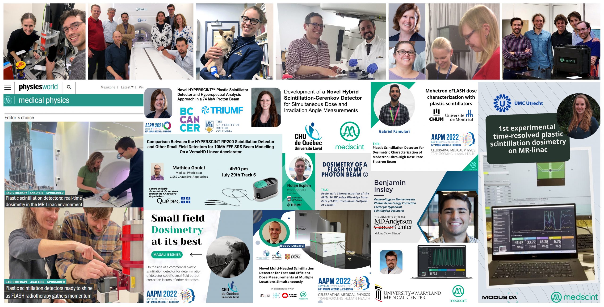
  • Comparison Between the HYPERSCINT RP200 Scintillation Detector and Other Small Field Detectors for 10MV FFF SRS Beam Modelling On a VersaHD Linear Accelerator

    Plastic scintillation detectors (PSDs) have advantageous dosimetric properties, including small size and energy independence, which make them ideal candidates for small field dosimetry.

    2021 AAPM ANNUAL MEETING
    J.Morin, JF.Cabana, M.Goulet, D.Theriault | CISSS – Chaudiere-Appalaches, Lévis, QC, CA
  • Characterization of the HYPERSCINT Dosimetry System for Real-Time Dosimetry Measurements with the Varian TrueBeamLinac

    Plastic scintillator/optical fibre dosimetry systems are advantageous due to their near water equivalence, waterproof construction, linear dose response, and good spatial resolution due to their small size. The nanosecond decay times of plastic scintillators enable the possibility of real-time dosimetry. We tested the new HYPERSCINT fibre detector system to determine if, in addition to the expected dose and field size responses, this system can provide real-time dose information. The HYPERSCINT system is suitable after appropriate calibration to be used to measure relative dose delivered in cGy as well as indicate changing dose conditions within 0.3 seconds.

    2020 AAPM AM
    C.Penner (1,2), C.Hoehr (2), C.Mendez (1), C.Duzenli (1) | BC Cancer, Vancouver, BC, CA, TRIUMF, Vancouver, BC, CA
  • Brachytherapy Technique Commissioning Using the HYPERSCINT Plastic Scintillation Detector

    Accurate dosimetry in brachytherapy is not an easy task, as most detectors exhibit volume averaging or energy dependence reducing their usability. Free from these limitations are plastic scintillation detectors, which makes them well suited for brachytherapy applications, either for in vivo dosimetry or commissioning. This work aims to determine if the HYPERSCINT scintillation dosimetry research platform can be used for brachytherapy dose measurement in the context of commissioning a new brachytherapy technique.

    2020 AAPM ANNUAL MEETING
    M.Goulet, N.Octave, P.Duguay-drouin | CISSS – Chaudiere-Appalaches, Lévis, QC, CA
  • Water-equivalent plastic scintillation detectors for high-energy beam dosimetry: II. Properties and measurements

    The properties of a new scintillation detector system for use in dosimetry of high-energy beams in radiotherapy have been measured. The most important properties of these detectors are their hgh spatial resolution and their nearly water-equivalence.

    PHYSICS IN MEDICINE & BIOLOGY
    A.S. Beddar, T.R. Mackie, F.H. Attix | Depanment of Medical Physics, University of Wisconsin Medical School, Madison, Wl, USA
  • Water-equivalent plastic scintillation detectors for high-energy beam dosimetry: I. Physical characteristics and theoretical consideration

    A minimally perturbing plastic scintillation detector has been developed for the dosimetry of high-energy beams in radiotherapy. The detector system consists of two identical parallel sets of radiation-resistant optical fibre bundles, each connected to independent photomultiplier tubes.

    PHYSICS IN MEDICINE & BIOLOGY
    A.S. Beddar, T.R. Mackie, F.H. Attix | Depanment of Medical Physics, University of Wisconsin Medical School, Madison, Wl, USA
  • Beam matching for small-field dosimetry applications using accelerator solenoid current and a miniature plastic scintillation detector.

    The goal of this work is to determine the optimal accelerator solenoid current (ASOL) that minimizes the spread of measured small field output factor (OF) values between a series of machines. Small field OF measurements of 6 MV flattening filtered beams from 3 Varian TrueBeam linear accelerators were performed using a 1 mm diameter by 1 mm length plastic scintillation detector HYPERSCINT HS-RP200 research platform. In conclusion Small field dosimetry characteristics are highly sensitive to beam focal spot size. and the possibility to optimize accelerator focusing coil current to reduce OF and penumbra width spread enables new avenues in beam matching of series of machines, especially for SRS and SBRT techniques.

    2022 COMP ANNUAL SCIENTIFIC MEETING
    L.Gingras (1), F.Beaulieu (1), M.Besnier (1), B.Côté (2), D.Leblanc (2), L.Beaulieu (1), L.Archambault (1) | 1- CHU de Québec – Université Laval, QC, CA, 2- MEDSCINT, QC, CA
  • Plastic Scintillation Detector for Dosimetric Characterization of Mobetron Ultra-High Dose Rate Electron Beam

    The purpose is to present commissioning data for the MOBETRON electron radiation therapy system (IntraOp) at ultra-high dose rate using the HYPERSCINT plastic scintillation detector. The suitability of using a plastic scintillator as an active dosimeter for commissioning measurements of an ultra-high dose rate electron beam has been demonstrated (reference dosimetry, DPP, beam penetration, linearity with number of pulses, linearity with PW and short-term output stability).

    2022 AAPM ANNUAL MEETING
    G.Famulari (1), K.Zerouali (1), O.Piron (1), JF.Aubry (1), F.DeBlois (1), JF.Carrier (2) | 1- Centre Hospitalier de l’Universite de Montreal (CHUM), Montreal, QC, CA, 2- Departement de Physique, Universite de Montreal, Montreal, QC, CA
  • Small field dosimetry of a Varian TrueBeam High Definition MLC linear accelerator using theHyperscint RP200 scintillation detector.

    Small field dosimetry of a Varian TrueBeam High Definition MLC linear accelerator using the Hyperscint RP200 scintillation detector.

    Purpose: To evaluate the performance of the new Hyperscint RP200 plastic scintillator for small field measurements of a Varian TrueBeam linear accelerator in comparison with the current state-of-the-art methodology in the clinic.

    Methods: Small field measurements were performed using different detectors: a diamond detector (microDiamond, PTW), a diode (Razor, IBA), a compact ion chamber (Razor, IBA), and a 1mm x 1mm
    plastic scintillation detector (Hyperscint RP200, Medscint). Correction factors based on measured field sizes, following TRS483 recommendations, were applied to all measurements. Output factors of a
    TrueBeam linear accelerator were measured for field sizes of 0.5 cm to 2 cm for jaws and MLC configurations for 6-MV, 6-MV FFF and 10-MV FFF photon beams. Output factors for different circular collimators (0.4 cm to 2 cm) were also obtained at 10-MV FFF. Scintillator measurements were compared to the small-field dosimetry methodology used clinically.

    Results: No correction factors were necessary for the plastic scintillation detector measurements. Scintillator measurements were within 1.1% of the standard methodology for all the small field geometries studied. Average relative differences were (0.3±0.5)%, (0.7±0.3)%, and (0.2±0.2)% for the 6-MV, 6-MV FFF, and 10-MV FFF, respectively. Output factors of circular field sizes down to 0.4-cm diameter were obtained with an average relative difference of (0.1±0.4)%, including a maximum difference of 0.7% for the smallest field.

    Conclusion: This new scintillation dosimetry research platform shows great promises for small field dosimetry. It has the potential to be used as part of a single-detector no-correction-factor methodology.

    2021 COMP ASM
    L.Gingras, B.Côté, F.Berthiaume, S.Lambert-Girard, D.Leblanc, L.Archambault, L.Beaulieu, F.Therriault-Proulx | CHU de Quebec – Universite Laval, QC, CA, Département de radio-oncologie et Axe Oncologie du CRCHU de Québec, QC, CA, MedScint, QC, CA